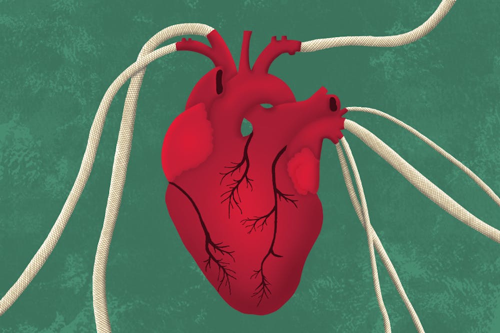
In his lab, Yuxiang Zhu leaned over a softly humming 3D printer, his face lit by the faint glow of a nearby monitor. Beneath the printer's nozzle, a thick blend of seaweed-derived sodium alginate and gelatin methacryloyl began to take shape. Layer by layer, an artificial blood vessel is printed to replicate the strength and stretch of the real thing.
Zhu, who recently earned his PhD in manufacturing engineering at ASU, played a central role in developing a 3D-printed vascular graft that behaves like a natural artery. This innovation could one day help reduce failure rates in bypass surgeries and lead to safer, more effective treatments for cardiovascular disease.
Zhu explained that the issue with existing grafts is that they cannot fully replicate how our blood vessels respond to stress and movement.
"That's the reason we want to introduce this structure. Because it matches the mechanical behavior with native blood better, so the failure rate will be lower," Zhu said.
The inspiration behind the graft's unique design came from a blend of biological insight and practical engineering. His adviser, Kenan Song, a former associate professor at ASU and now at the University of Georgia, shared how the structure was influenced by nature itself.
"If you take a look at the structure (of blood vessels)... each layer will function differently," Song said. "However, if you look at the manually made materials, they usually have mismatched properties with the human blood vessels."
That gap inspired the team to create a multilayered, flower-mimicking design. The interior is solid, the exterior is solid with a hollow structure in between. The cross-section of the 3D prints resembles a simple drawing of a daisy-like flower.
"When it's filled with blood, it will cause pressure and eventually the exterior layer will protect the interior layers," Song said.
This structure allows the graft to stretch and adapt, just like a natural artery. More importantly, it narrows the performance gap between manually engineered materials and real human vessels.
"It makes the materials we human beings make behave more similarly to the human being artery systems," Song said.
The research team ran both experiments and computer simulations to validate their approach.
"To predict if it would last 20 years, you probably don't want to do the test within 20 years. It's almost one-fifth of your lifetime, right? So we'll use modern simulation to predict behavior that would be experimentally challenging," Song said.
Ensuring biocompatibility was another critical milestone. Biocompatibility means the new materials do not harm or are not rejected by human cells. Yu Shrike Zhang, an associate professor at Harvard Medical School and a collaborator on the project, led cell viability tests to ensure the printed grafts wouldn't be toxic to living cells.
"Getting the cells into these tiny tubes is always something that's a little challenging. But once the cells are in there, they looked pretty happy in general," Zhang said. "But then, if you want to use this as a like implant, especially within human system, they have to be staying stable over time. They cannot rupture."
The long-term goal is to move the grafts from lab to clinic, but it won't happen overnight.
"We have had a very good first step," Song said. "We are still looking for parts to make the material and structure more consistent. It's about the quality control."
Such grafts could also have real-world applications beyond hospitals.
"We want to provide timely and efficient curing on the battlefield. So how do we solve that?... You need decentralized solutions. With 3D printing, we can create on-demand grafts in those kinds of urgent situations," Song said.
With teams like this combining nature-inspired design, modern engineering and an eye toward real-world use, the future of regenerative medicine is not being imagined— it's being printed.
Edited by Sophia Ramirez, Abigail Beck and Natalia Jarrett.
Reach the reporter at ammaarzindani@asu.edu.
Like The State Press on Facebook and follow @statepress on X.