ASU researchers have partnered with other schools and health institutions to provide a breakthrough in CTE diagnosis, using an experimental imaging method to identify the condition in living patients.
The study, which was released earlier this month in The New England Journal of Medicine, aims to help solve a problem in treating Chronic Traumatic Encephalopathy (CTE), which is only diagnosable after death when doctors have access to a patient's brain tissue through an autopsy.
While the type of PET scan used in the study is still experimental and not currently being used for clinical purposes, it may provide a pathway toward one day diagnosing CTE in living patients, said Dr. Charles Adler, neurologist at the Mayo Clinic in Phoenix.
"We're trying to find a way to diagnose CTE in living patents and living individuals," Adler said. "So this is just the first step."
CTE is a neurodegenerative disease that spreads through the brain, slowly kills brain cells and shrinks brain mass. This disease can lead to many behaviors that range from memory loss to aggression and even dementia.
CTE falls into a category of neurodegenerative diseases known as tauopathies. Tauopathies are characterized by the abnormal buildup of the tau protein in the brain. Some other conditions that also fall into this category include frontotemporal dementia, Alzheimer’s disease and post-traumatic stress disorder.
Along with the buildup of protein, CTE involves head trauma, a feature not seen in other tauopathies. That in itself is an issue that makes it difficult to identify.
The accumulation of tau protein cannot be seen using traditional imaging methods, like MRI or CT scans, making it difficult to diagnose someone with multiple concussions and clear symptoms with CTE.
However, with the new study, researchers have found that they can use an experimental imaging method, called a flortaucipir PET scan, to screen for tau buildup in the brain.
CTE has received a lot of attention in recent years for being linked to a slew of deaths in former NFL players.
A 2017 study done by the Boston University School of Medicine found that 99% of the 111 deceased former NFL player's brains observed in the study had Chronic Traumatic Encephalopathy, better known as CTE.
Dr. Eric Reiman, a professor of neuroscience at ASU and executive director of the Banner Alzheimer’s Institute, has done extensive work in brain imaging research and said that CTE presents unique challenges compared to other neurodegenerative diseases.
Unlike other medical conditions, there are no confirmed symptoms that CTE exhibits in living patients, Reiman said, forcing doctors to rely on other biological factors that exist in patients' brains to estimate the likelihood that they have CTE.
“CTE is defined based on biology, not in symptoms yet," he said.
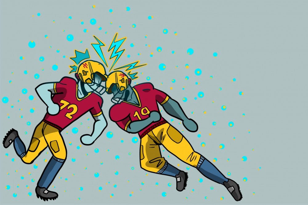
CTE is found most commonly in football players, but it occurs in anyone who has suffered from repetitive hits to the head, which can include boxers and combat veterans. These hits don’t necessarily have to be large, concussive hits, but rather an accumulation of hits over time that can lead to the buildup of the tau protein.
Diego Mastroeni, an assistant research professor at the ASU-Banner Neurodegenerative Disease Research Center, said he initiated the research partnership on CTE diagnosis after personally hearing concerns about CTE from a former football player.
“I was contacted by my father in law’s cousin who had played in the NFL for many years and was having some cognitive problems, memory problems and some other problems as well,” Mastroeni said.
With this in mind, Mastroeni said he gathered a team including local researchers from ASU, Banner Alzheimer’s Institute, Mayo Clinic College of Medicine, Brigham and Women’s Hospital and Boston University to experiment with the flortaucipir PET scan.
The study was conducted under the Diagnostics, Imaging And Genetics Network for the Objective Study and Evaluation of CTE (DIAGNOSE CTE) research project, which is funded by the National Institutes of Health to "collect and analyze neuroimaging and fluid biomarkers for the detection of CTE during life," among other goals.
In the study, researchers imaged a group of former football players and compared that to images they took of people who also suffered from similar symptoms, but did not experience any head trauma. The brains of the football players showed significant amount of tau. It was concluded that they most likely had CTE due to the fact that they lacked the other main physiological signs that are typically seen in other tauopathies.
“At the time, we didn't have the tau scan that we're doing now, we didn't have that available, so we weren't able to give (my father in law's cousin) a real clinical diagnosis," said Mastroeni. "The reality is we didn't know what was going on.”
Although the study represents a breakthrough in the realm of diagnosing CTE, Reiman said there is a lot of work yet to be done.
“We can not use these biological techniques to confirm the diagnosis of CTE or explain a person's memory and thinking problems," he said. "But we think this provides a foundation for making progress towards that end.”
According to Adler, more research is already on the way at the DIAGNOSE CTE project.
“We're in the process of enrolling 120 NFL players, 60 former NFL players, and 60 former college players," he said. "And as part of that study, we’re doing the experimental pet scans, we’re doing spinal taps and we're checking blood. So we're looking for (more) biomarkers to suggests that these individuals have, or may have CTE”.
While there is still yet to be a way to diagnose CTE in living people, the recent study hopefully serves as the first step in leading more research in the future.
The flortaucipir PET scan could also be the key to helping people with CTE by being used to determine a patient's level of treatment by a doctor based on the amount of tau in the brain. In the future, this technique could also be used to detect if certain populations could be more susceptible to developing this disease.
Reiman said he is hopeful that the future of CTE diagnosis will advance quickly and that "we will see significant progress in two to three years.”
Until then, he said that preventative measure such as padding NFL helmets and avoiding activities that may lead to head trauma can help lower the risk of developing this disease in the first place.
Reach the reporter at rwondimu@asu.edu or follow @_beccaswan on Twitter.
Like The State Press on Facebook and follow @statepress on Twitter.


